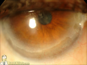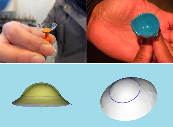

Usually, the entire cornea is clear, and no vascularization, Fleischer ring, lipid deposits, or changes in corneal sensitivity are observed. Rarely on presentation, these patients can report a sudden, painful red eye with watering accompanied by a sudden drop in vision and photophobia due to acute hydrops and/or spontaneous corneal perforation.Įxamination reveals stromal thinning of the periphery of the cornea, extending mostly from 4 o'clock to 8 o'clock, in a crescent shape, and has a slow and progressive development. The high against-the-rule astigmatism stems from a paradoxical steepening at 90 degrees. The steepening and ectasia occur at the junction of affected and unaffected tissue, leading to the typical high cylindrical loop. The flattening in the vertical meridian is due to tissue loss and a thin stroma in a crescentic pattern. On evaluation, high irregular, against-the-rule astigmatism is commonly found. But best-corrected visual acuity becomes poor only in advanced stages. Most commonly, a gradual, progressive diminution in vision or longstanding poor visual quality is the presenting feature of these patients. Coexistent retinitis pigmentosa, chronic open-angle glaucoma, scleroderma, retinal lattice degeneration, eczema, and hyperthyroidism have been observed in some patients with PMCD, but no direct causative association has been found to be true. Also, floppy eyelid syndrome found in obese patients may lead to chronic mechanical rubbing of the cornea leading to thinning and ectasia.Ītopic and vernal keratoconjunctivitis has been linked in reports to superior PMCD. The repeated occurrence of hypoxic conditions in these patients may be hypothesized to lead to anaerobic glycolysis and stromal acidosis, and in turn, promote transcription of proinflammatory cytokines, tumor necrosis factor-alpha or interleukin-6, leading to corneal thinning.


Obesity and obstructive sleep apnea have been associated with PMCD in the literature. Similar to keratoconus, electron microscopy of the cornea in PMCD reveals abnormally spaced collagen fibers with a periodicity of 100 nm to 110 nm, as opposed to 60 nm to 64 nm found in normal corneas.


 0 kommentar(er)
